Interesting article in The Horse about Daisy Bicking regarding rehabilitating horses with Equine Metabolic Syndrome related laminitis. She has been getting incredible results with the protocols she has developed. I have taken a couple of workshops with Daisy and look forward to taking more to learn more about helping these horses. Take a look at the full article
Long Toes and Muscle Pain
An interesting article in The Horses showing the connection between long toes and pain in the gluteal muscles. The researchers have shown that by reducing the breakover in the hind toes of a horse, it can reduce the pain reaction to palpation in the gluteal muscles.
Importance of X-rays in Founder Trims
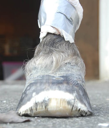
Last month I spent some time down in Pennsylvania at Daisy Haven Farm doing come continuing education learning the value of having x-rays to more accurately trim a horse’s hoof. Part of the workshop was a day of looking at hoof distortion and how to correct for it. To aid in this, cadaver hooves are x-rayed before they are trimmed and then again after the trim. Later the group gets together to critique the effectiveness of the trim. Since I had done Daisy’s 5 day workshop on this in May, I knew the procedure, so I found the most foundered cadaver foot available to improve my eye for trimming for that type of distortion.
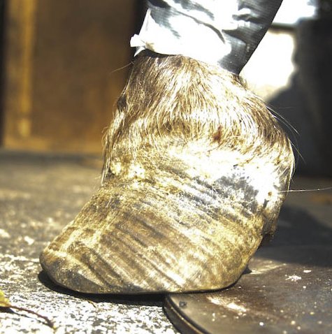 This foot had quite a severe dish and is quite long.
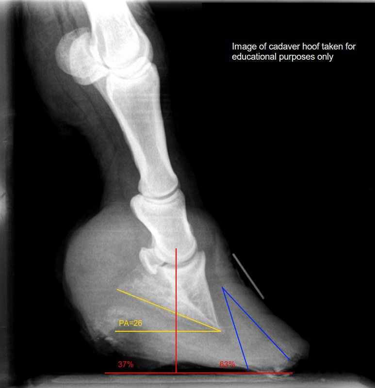
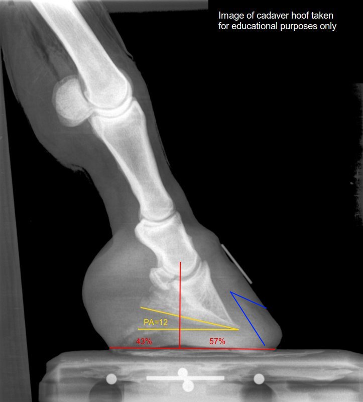
Looking at the x-ray, the coffin bone ( the triangular bone in the hoof capsule) has rotated quite a bit away from parallel to the hoof wall, which is where it normally should be located. The bottom part of the coffin bone should be at have a 3-8° angle with a line parallel with the ground. This is called the palmer angle (PA). In this x-ray, the palmer angle is 26 and is shown in yellow. The blue lines show the lamellar wedge, which is made up of stretched lamella (the material that attaches the hoof to the coffin bone) and poorly organized hoof material. This is the material that fills the gap that exists between where the hoof wall is and where it should be. The Center of Rotation (COR) is the point at the end of the second paster bone (P2) around which the coffin bone rotates. It has been found that in a healthy foot, half of the foot will be in front of a line dropped down from the COR and half will be behind it. The red lines show the balance of the hoof around the center of rotation . The COR is at the top of the vertical red line.
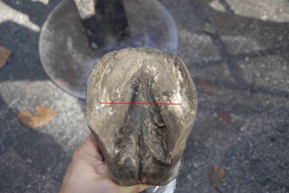 On the sole of the foot, a line projected down from the COR usually corresponds to landmarks on the sole, such as the widest part of the sole, the end of the bars and the bar swells (not clear on this photo). It is usually about an inch behind the apex of the frog. The red line on the photo of the sole of the foot has been placed using measurements from the x-ray, and is quite close to where it would be placed using the landmarks on the sole of the foot, showing the reliability of those landmarks.

The toe of the foot before trimming shows a considerable dish. The top 1/2 to 3/4 inch of the hoof wall beneath the coronary band (shown by the green line) gives an indication of where the wall would be growing if the wall was well connected. This angle is used to guide the removal of material from the toe wall.
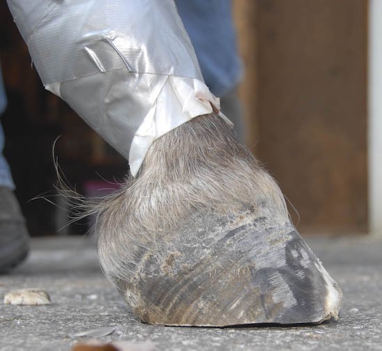
After the center of the foot was determined, the heels were shortened to bring the coffin bone back to a more normal angle and the lamellar wedge was reduced using a rasp and a belt sander. This brought the toe to an angle that approximates where it would be if the wall was still well connected. This will expose the lamellar wedge, but it is sufficiently hardened that it will provide sufficient protection for the sensitive lamina. The toe was then bevelled to bring the balance of the foot closer to the ideal 50/50 ratio, resulting in a foot that looks much closer to normal
After trimming another x-ray was taken and as you can see, the palmer angle has been reduced, and is much closer to the ideal 3-8°. The balance of the foot is much closer to the ideal. There is still some heel that could be removed and that would bring the foot closer to the ideal palmer angle, as well as improve the ratio around the COR.
This shows the importance of having quality x-rays of the foot, so that it is possible to get a better idea of the angles of the internal structures within the foot before attempting to improve the hoof. The external markers are very useful, but more rapid rehabilitation is possible through the use of x-rays.
Farriers Appreciation Week
Thank you to Joanna Kirby for her kind words submitted to American Farriers Journal for Farriers Appreciation week. I am very glad that I have been able to help Chelsea with her hoofcare.
Take a look at what has been said about other farriers, including one of my instructors and mentors Daisy Bicking on the American Farriers Journal Website
Recent Workshop at Daisy Haven Farm
At the 2013 International Hoof Care Summit, I attended a talk by Daisy Bicking of Daisy Haven Farm. In her talk she showed how she has had an over 90% success rate at treating founder. At the conference I decided that I needed to find out more about her methods and so asked about workshops.
In early May, I attended a 5 day workshop at Daisy Haven Farm on hoof deformation. In this workshop, 60 cadaver hooves were x-rayed, and then after Daisy did a demonstration of her method of hoof mapping, we chose a hoof to work on. After we did our trim, the hooves were re-x-rayed and in the afternoon we got together and did a critique of each other’s trims. It was an eye opening thing to see how much could be taken off a foot before you got into the sensitive part of the foot. On the third day, Paige Poss, one of the early barefoot trimmers did a dissection of several of the feet that had been trimmed, giving the attendees a much better feel for how the hoof is constructed. On the forth day of the workshop, we went to a local horse rescue and attended to some of the horses there, and had a further chance to have our work critiqued by Daisy. The last day was on how to apply composite shoes for more severe cases of founder.
It was an incredible learning experience that has given me a number of new tools to use in the treatment of founder and hoof deformation in general, and I feel has taken my skills to a new level. An added benefit of the workshop is that I was invited to a closed Facebook group of students that have been to Daisy’s workshops, which has given me access to over 90 farriers and veterinarians with which I can discuss difficult and unusual cases.
For those on Facebook, a number of photos from the workshop are available here.
Veterinarian Reviews the Barefoot Concept
There is an interesting article in The Horse in which Debra Taylor DVM discusses the advantages of the barefoot concept. Take a look at the full article here.
Research Showing the Benefits of Barefoot Trimming
An interesting article in The Horse discusses the findings of Hilary Clayton of Michigan State University shows that a technique that leveled the hoof to the live sole, lowered the heels, beveled the toe, and rounded the peripheral wall where the sole, frog, and bars were left intact was beneficial to the horse and was over time able to correct under run heels. It also showed that the frog, bars and sole should play a part in weight bearing. The whole article can be read on The Horse website
Barefoot More Accepted In High Level Competition Horses
Shannon Peters, wife of Steffen Peters, has taken her Grand Prix horse barefoot, along with Ravel, Steffen’s Olympic horse, who is now retired. A complete article on this is available at the Dressage Today website
Start With The Beginning

Various Coffin Bones
The coffin bone, also known as the pedal bone or the third phalanx is totally enclosed in the hoof capsule and is what what controls the shape of a horse’s hoof. In horses that have a short wide coffin bone, such as the one in the upper left in the photo will have a foot that is wide and short, which is quite common in Thoroughbreds. Other horses will have coffin bones that are long and narrow, such as the one on the upper right, giving a lon narrow foot. Also note that the coffin bone has a number of holes in the surface. This is where blood vessels are located in the bone and it is through these vessels that the laminae are nourished. The bottom of the coffin bone is convex and the sole of the foot follows this shape to form the bottom of the foot. The coffin bone is articulated with both the navicular bone and the short pastern bone. This is in the form of a hinge joint that allows the foot to bend back and forth, but has little side to side movement. At the back of the coffin bone are the wings of the coffin bone. These are attached to the lateral cartilages, and in some cases the lateral cartilage will change to bone (ossify) and create what is known as sidebone. This can be seen on the right side of the coffin bone on the bottom right. The point at the top of the coffin bone is the attachment point for the digital extensor tendon, and the deep digital flexor tendon inserts on the bottom of the coffin bone along what is called the semi-lunar crest.
Barefoot Benefits
There are a number of benefits to a horse going barefoot. The most obvious is that there is less damage to the hoof capsule from the use of nails. Nail holes will weaken the hoof wall and create an environment for anaerobic bacteria and fungus to get a foothold in the hoof wall and possibly cause problems later on, such as white line disease.
The greatest advantage to the horse is that he will have better proprioception, that is, he will have a better sense of where his foot is, and what is underneath it. A horse in shoes is like you going around wearing heavy gloves all the time. You can still get a general feel for something, but there is a lack of sensitivity. With better proprioception, the horse will be more surefooted and have a better sense of his environment and can make compensations and adjustments for uneven or slippery footing.
In terms of long term health, if a horse is left barefoot, he will develop a better internal structure within the hoof and is likely to stay sound longer. The effect of a shoe on a foot is similar to putting a cast on your arm. Since the structure cannot move as it was intended, it will tend to atrophy, much as the muscles in your arm will atrophy due to a cast. The most damage is done when a horse is shod at an early age, as are many racehorses. This will not allow the internal structures of the foot to develop while the animal is growing, and in some cases will cause the animal to permanently have poor quality feet.
A further advantage is for the social health of the horse. If a horse is shod, particularly on the hind feet, it is dangerous to turn them out with other horses, due to the possibility of an injury due to a kick. When barefoot, horses can be turned out in groups which is much better for their social health, as horses are herd animals and generally do better when they are allowed to interact with other horses.